Juan Caicedo
As described in our lab’s recent review article, there are so many great reasons to use microscopy images to create signatures of perturbations: identifying phenotypes associated with disease, identifying chemical mechanisms of action, and discovering gene functions, among others. Have you wanted to give this approach a try, but been overwhelmed by the computational options available?
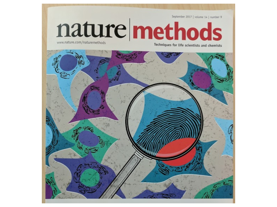
Our recent paper, Data analysis strategies for image-based cell profiling, could come to the rescue. This Nature Methods paper is a comprehensive survey of computational methods and techniques that several labs around the world have found useful for extracting quantitative information from images. Our lab was one of the more than 20 that participated in this effort, which inspired the cover of the journal in the September 2017 issue.

When researchers use images to objectively quantify cellular states, there is a lot of information that can be extracted from what appears to be unstructured images. That information is processed in multiple sequential steps that involve transforming and aggregating data from one stage to the next. At each stage, computational biologists may face several decisions, such as, “what method should I use to get the right output”, and “how do I even know I’m measuring the right signal?” These questions are addressed in the review paper by enumerating each of the steps and making suggestions and recommendations for other researchers to follow what we consider to be the best practices known in the image-based cell profiling (or “morphological profiling”) field.
The paper began in Boston during the summer of 2016, when more than 20 labs from around the world gathered together to share and discuss their pipelines. This was the first time a group of researchers met to discuss topics related to data processing methods for image-based cell profiling in a hackathon-like event. We agreed on the common steps to successfully run a cell profiling project using imaging. These eight steps are depicted in Figure 1 of the paper, and include everything from image processing to downstream statistical analysis.
As we are very enthusiastic about open and reproducible science, we also dedicate a section of the paper to discuss and make recommendations for sharing data and code. This is an important aspect to make progress in the field and realize the potential of imaging technologies as a powerful tool for studying biological systems. Therefore, we couldn’t be happier that Nature Methods agreed to publish the paper open access. Finally, we also discuss opportunities for new methods to improve the quality and accuracy of cellular profiling, especially deep learning and artificial intelligence as transformative technologies for image analysis.
We hope that the paper will be useful for anyone in the scientific community interested in extracting the most out of their images. We look forward to seeing the new biological discoveries made possible by microscopes, images and data analysis.
